How Do Motor Cortex Pathways Change in Aging?

Post by Rebecca Glisson

The takeaway

As we age, we sometimes lose our ability to move normally, which also significantly lowers our quality of life and capacity for independence. The motor cortex of the brain, which controls our movement, and the brain pathways descending from this area deteriorate in older adults, suggesting a focus on these areas could help us better treat neurodegenerative diseases.

What's the science?

Our movement is controlled by an area of the brain called the motor cortex, which can lose its function over time as we age. The motor cortex sends signals through two tracts, or pathways, of cells: the corticospinal tract (CST) and the corticostriatal tract (CStrT). This week in NeuroImage, Wen and colleagues investigated the changes in the CStrT that occur as people age, aiming better to understand the neural basis of movement-related neurodegenerative diseases.

How did they do it?

The authors wanted to study how aging affects several parts of the motor cortex and its pathways: its overall structure, how blood flows through this area, and the quality of the cells in these areas. To study these variables related to movement abilities, the authors used structural magnetic resonance imaging (MRI) to image and analyze the motor cortex in the brain. They also used diffusion MRI to analyze the pathways of the CST and CStrT. Linking this to movement, the authors measured participants’ motor function using endurance and locomotion walking tests and grip strength tests. The authors grouped participants into two age groups: the younger group, 36 to 65 years old, and the older group, 66 to 90 years old. 

What did they find?

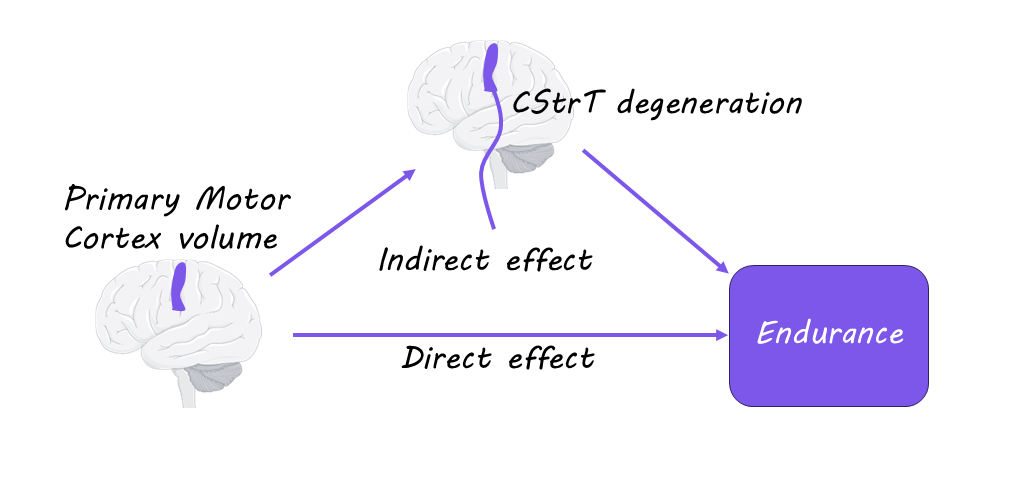
While younger participants had normal brain structure, the authors found that older participants had less volume in the motor cortex and less blood flow to this region. The older group of participants also had significantly lower movement abilities involving locomotion, endurance, and strength. This suggests that the loss of certain movement abilities as we age is due to degeneration of the motor cortex. The authors also found that the CST and CStrT pathways were of significantly lower quality in the older group of participants and that this deterioration mediated the relationship between motor cortex atrophy and decline in motor function. Therefore, the CST and CStrT pathways are particularly important to movement function and are affected in aging.

What's the impact?

This study is the first to show that the changes in the motor cortex and its related pathways in the brain due to aging are directly related to a loss of movement functioning in older adults. This highlights the need to focus on these areas for studying movement diseases related to aging. Studies like these can help us detect neurodegenerative disorders earlier and develop better and more effective treatments.

Access the original scientific publication here. 

Cyclical Brain Rhythms Drive Key Cognitive Functions

Post by Soumilee Chaudhuri

The takeaway

Brain networks carry out day-to-day cognitive functions, such as focusing, remembering, and processing sensory information. The activity in these networks follows a cyclical pattern, with each activation supporting essential cognitive functions.

What's the science?

The human brain carries out numerous cognitive and bodily functions, but it has been unclear how these processes are coordinated over time. Previous studies using different forms of neuroimaging have observed directional relationships in network transitions; however, it remained unclear whether these asymmetries are part of a higher-level organization. This week in Nature Neuroscience, van Es and colleagues analyzed brain imaging data from five independent datasets and revealed that, while individual transitions appear noisy, they collectively form consistent cycles that repeat every 300–1,000 milliseconds, with each network having a preferred position within the cycle

How did they do it?

The study analyzed brain activity from over 800 participants across five Magnetoencephalography (MEG) datasets. Participants ranged in age from 19 to 88 years and included both males and females. MEG signals were mapped onto the brain’s cortex, divided into 38–78 regions depending on the dataset, to track how different brain areas interacted over time. Using a Hidden Markov Model, the researchers identified 12 recurring brain network states and examined the timing and direction of transitions between these states. They applied a new method, temporal interval network density analysis or TINDA, to detect characteristic sequences of network activations emerging as cycles. The makeup of this cyclical pattern, along with its strength (amount of deviations from the cycle) and speed, were quantified and analyzed for consistency across participants and their relationship with age, cognitive performance, and behavior, showing how these brain rhythms relate to key individual traits.

What did they find?

Identified brain networks followed a cyclical pattern, with cognitive and perceptual networks taking turns in a consistent and coordinated sequence. This sequence or cycle lasted approximately 300–1,000 milliseconds, and the timing and strength of these cycles were linked to age and cognitive performance. The researchers found that older adults showed much slower cycles compared to younger participants. The phase of the cycle also predicted moment-to-moment behavior, including markers of memory consolidation and reaction speed. Finally, additional findings suggested that the rate of these cycles was partly heritable, indicating a biological basis for these rhythms.

What's the impact?

This study provides compelling evidence that the natural cyclical activation of large-scale brain networks underlies essential cognitive functions. These findings shed light on the mechanisms underlying cognitive processes in the brain and highlight the potential for targeting brain network rhythms in interventions designed to enhance cognitive function.

Access the original scientific publication here.

How Does the Brain Overcome Social Exclusion?

Post by Rebecca Glisson

The takeaway

Negative emotions that come from social exclusion can be relieved through emotionally supportive social interaction between two people. During this emotional support, there is synchronized activity in the prefrontal cortex, the brain region involved in emotional regulation, between the person giving and receiving emotional support.

What's the science?

When you are excluded from a social group, it can be challenging to manage the unhappiness and upset on your own and to emotionally regulate. Having someone else to help support and comfort you, which is called interpersonal emotional regulation, may be a more effective way to handle these negative emotions, although this has not yet been studied. This week in Scientific Reports, Zhu and colleagues investigated whether interpersonal emotional regulation is more effective than intrapersonal emotional regulation for managing negative emotions in response to social exclusion, as well as the brain activity that controls these behaviors.

How did they do it?

To study the differences between intrapersonal and interpersonal emotional regulation, the authors had participants experiencing negative emotions after social exclusion events either regulate their own emotions or work with another participant to regulate their emotions. First, participants were shown pictures of someone being excluded by their peers, and then were asked to think about a time they personally experienced social exclusion. Following this, one group of participants was given a strategy to try to regulate their emotions while alone, which simulated intrapersonal emotional regulation. The interpersonal emotional regulation group, on the other hand, was paired with another person who was given a strategy to help the person experiencing social exclusion.

In a second experiment, the authors used functional near-infrared spectroscopy (fNIRS) to measure brain activity while participants were negatively reacting to social exclusion and during the interpersonal emotional regulation afterwards. The authors focused their measurements on the prefrontal cortex, the area of the brain that links emotion regulation and social interaction. Both the participants who were experiencing the negative emotions and their paired partners who were helping regulate their emotions were scanned at the same time to study if brain activity was synchronized during interpersonal emotional regulation.

What did they find?

In the first experiment, the authors found that people feeling negatively after social exclusion were better at managing those emotions with someone else than by themselves. This suggests that interpersonal emotional regulation is more effective at relieving negative feelings after social exclusion than working through the emotions alone. In the second experiment, the authors found that the left medial part of the prefrontal cortex is more active while someone is reacting negatively to social exclusion. Then, during interpersonal emotional regulation, they found that the activation of both the emotion experiencer and the emotion supporter was synchronized in the prefrontal cortex. This suggests that synchronization of activity in the prefrontal cortex between two people during interpersonal emotional regulation is the mechanism that leads to better outcomes after social exclusion.

What's the impact?

This study is the first to show that interpersonal emotional regulation is more effective than intrapersonal emotional regulation at reducing negative reactions to social exclusion, and the brain mechanisms involved with this process. These results suggest that empathy is crucial for helping others deal with social exclusion. As social exclusion is common for both children in school and adults in the workplace, and can lead to poor outcomes for both mental and physical health, it is important for studies like these to provide strategies to manage responses to social exclusion.

Access the original scientific publication here.